Bacterial OMVs are spherical membrane structures, typically 50–250 nm in diameter, released during bacterial growth. They carry lipopolysaccharides, peptidoglycan, membrane and cytosolic components, proteins, toxins and nucleic acids, influencing virulence, horizontal gene transfer, phage infection, metabolite transport and bacterial interactions. OMVs have been used as vaccine adjuvants. Their lipid-and-protein membranes resemble cell membranes and offer potential as drug delivery carriers.
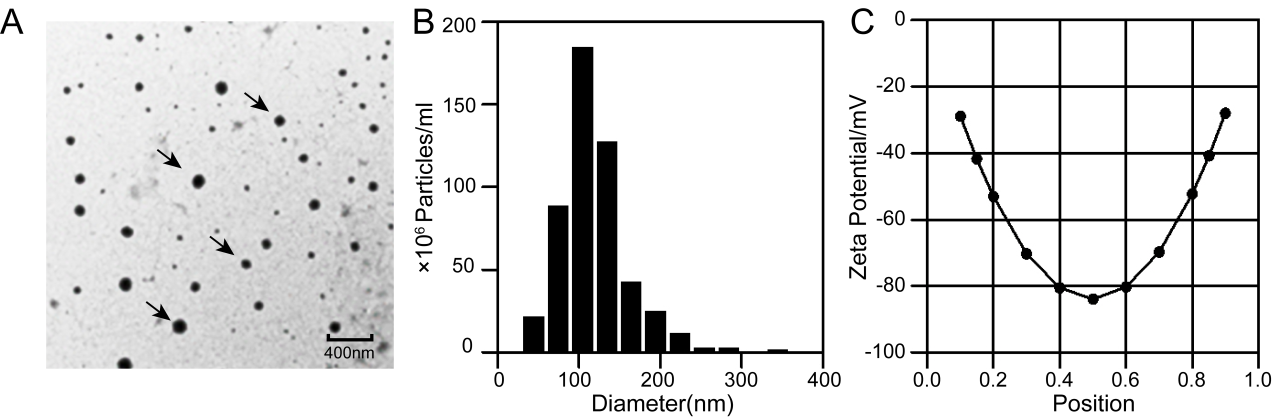
Figure 1. Properties of the OMV Samples
The team characterized OMV morphology, particle size and surface potential, confirming structurally intact, stable vesicles.
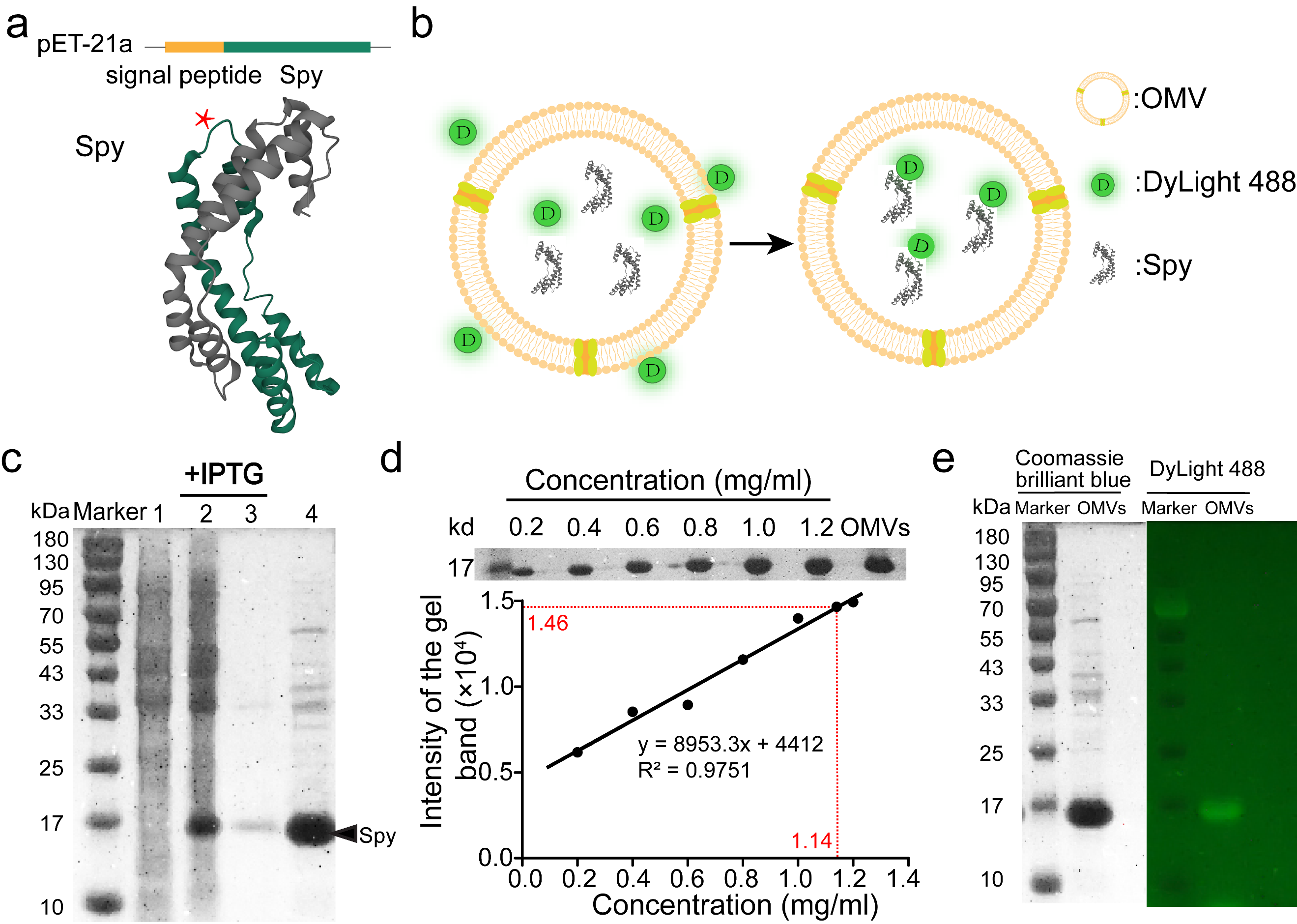
Figure 2. Enrichment and Labeling of Target Proteins Inside OMVs
To load target proteins into OMVs, the team constructed a secretory expression plasmid (Figure 2a),directing Spy to the periplasm for packaging into OMVs. Protein analysis showed enrichment up to 62 mg/mL (Figure 2c,d). Fluorescent probes that pass across the vesicle membrane successfully labeled the encapsulated protein (Figure 2e).
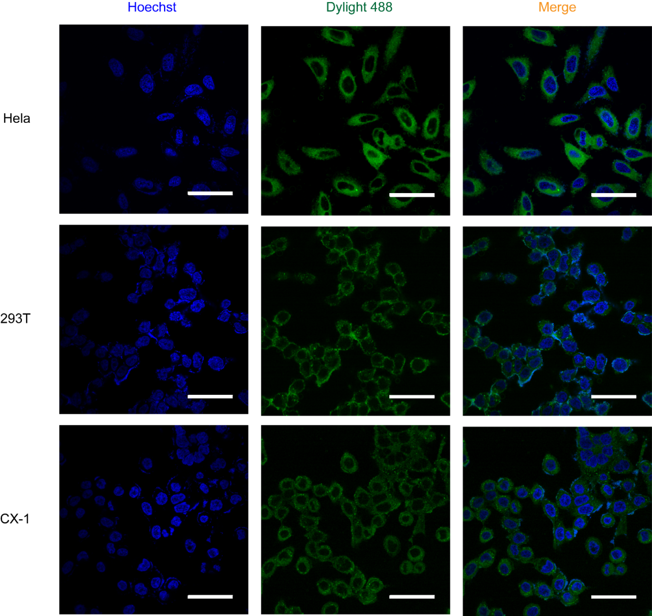
Figure 3. CLSM Images of Different Cell Lines Treated with OMVs Containing Labeled Spy T72C
Confocal microscopy showed that simple incubation with OMVs delivered target proteins into the cytosol of multiple cell lines, with delivery efficiencies of approximately 90% (Figure 3).
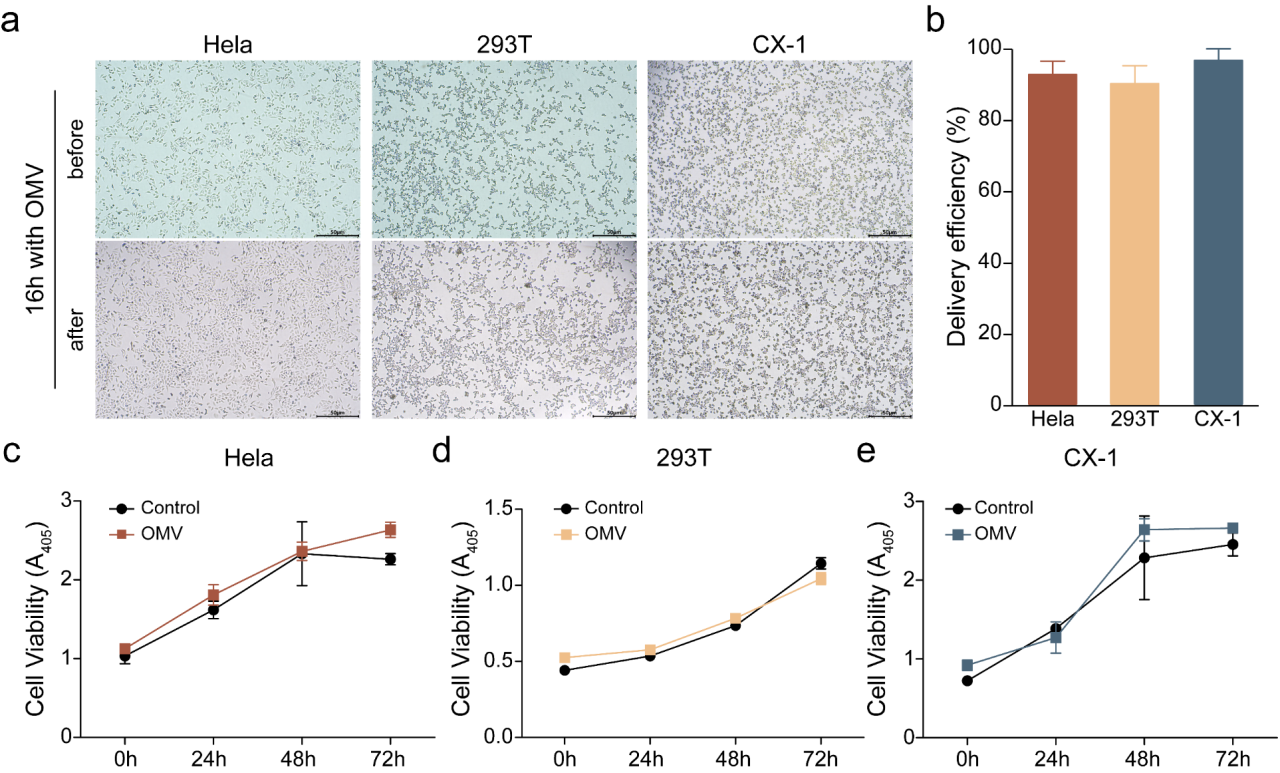
Figure 4. Confocal Microscopy Results
An initial cytotoxicity assessment found no apparent effect of OMVs on normal cell growth over 72 hours (Figure 4).
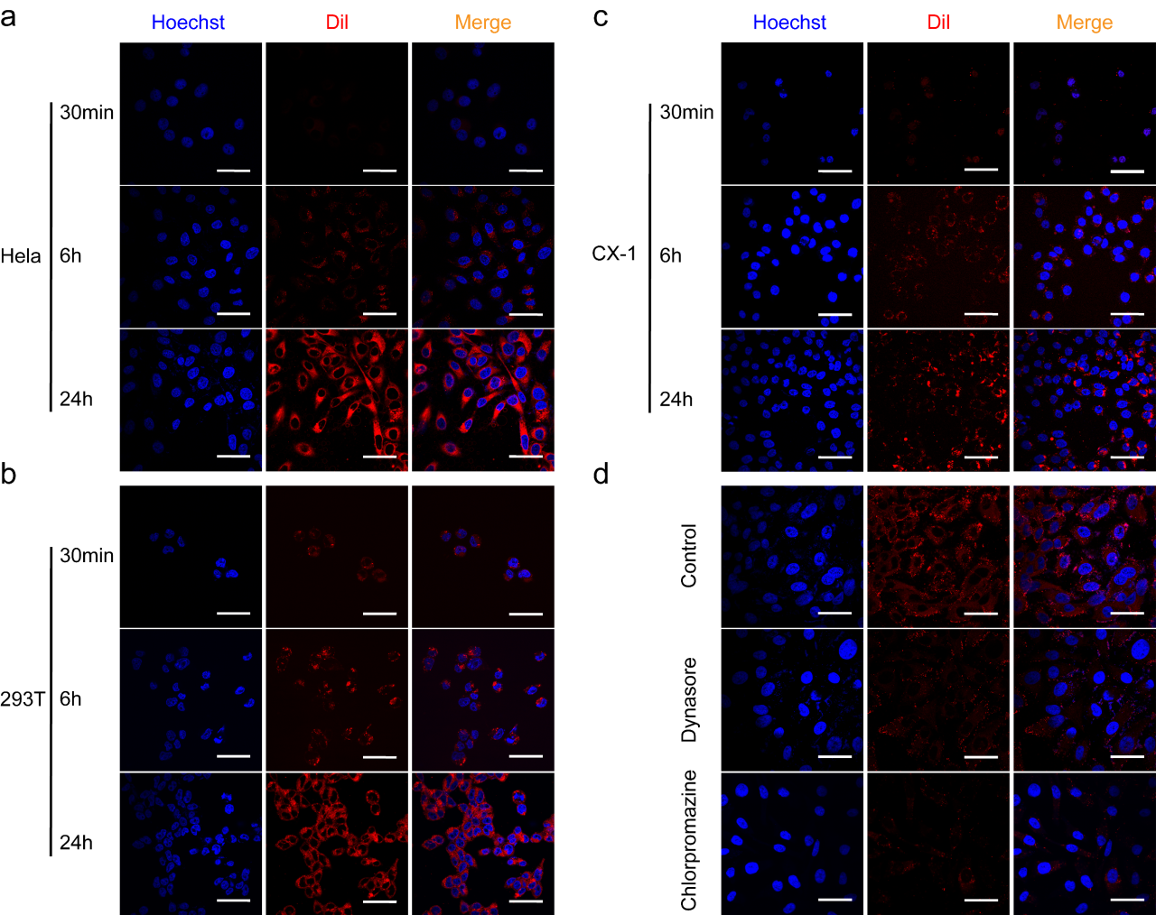
The team also investigated delivery mechanisms of E. coli-derived OMVs and found that uptake across multiple cell types occurred through endocytosis (Figure 5).
Figure 5. Incubation with DiI-Labeled OMVs
The authors also investigated delivery mechanisms of E. coli-derived OMVs and confirmed endocytic uptake in multiple cell types (Figure 5).
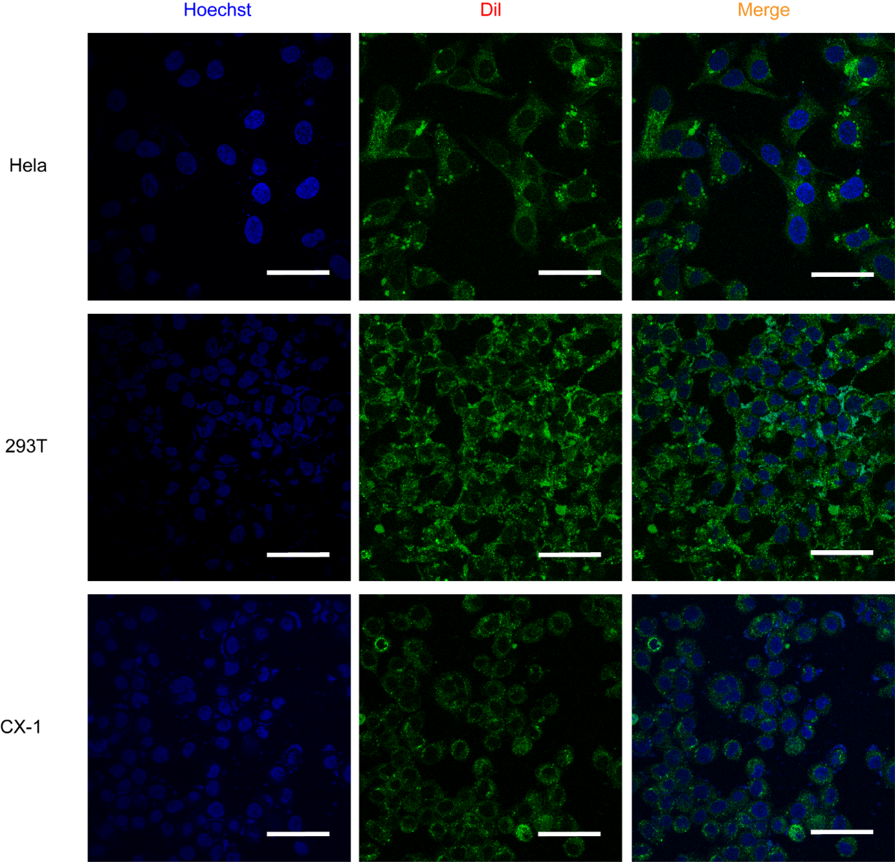
Figure 6. OMV-Mediated Delivery of Labeled Nanobodies to Different Cell Lines
Confocal images of HeLa, 293T and CX-1 cells after 12 h incubation with OMVs carrying labeled nanobodies. Scale bar: 50 μm.
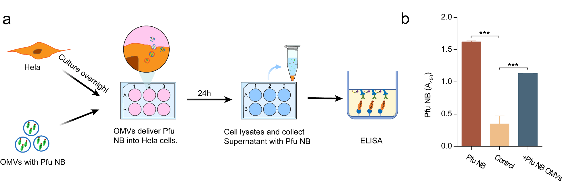
Nanobodies were packaged into OMVs using the same approach. Confocal microscopy supported nanobody delivery through this system (Figure 6), and ELISA further showed that the delivered nanobodies retained biological activity (Figure 7).
Figure 7. Biological Activity of Pfu Nanobodies Following OMV-Mediated Delivery
Together, these experiments demonstrate efficient enrichment, specific labeling and cytosolic delivery of target proteins using an OMV-based intracellular delivery system.